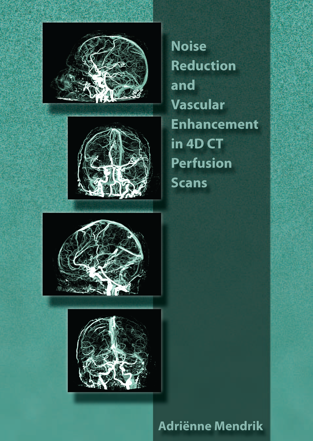
Noise Reduction and Vascular Enhancement in 4D CT Perfusion Scans
A. Mendrik
- Promotor: M. Viergever and W. Prokop
- Copromotor: B. van Ginneken and E. Vonken
- Graduation year: 2010
- Utrecht University
Abstract
Computed tomography (CT) uses X-ray radiation to construct images. Applying X-ray radiation to the human body may damage the tissue and increases the risk of inducing cancer. Therefore, the radiation dose should be kept as low as reasonably achievable (ALARA). This is especially true for 4D CT perfusion (CTP) scans, since these scans consist of multiple sequential 3D CT scans over time. Therefore, not much radiation dose can be used per sequential scan. However, lowering the radiation dose increases the amount of noise in CT scans and therefore decreases image quality. This thesis describes two ways of dealing with radiation dose reduction in terms of image processing: 1) Image processing techniques can be utilized to extract as much information as possible from one CT scan, obviating the need for additional CT scans, therefore reducing radiation dose. Chapter 2 describes a method to automatically derive vascular information from 4D CTP scans and separate the arteries from the veins. The resulting CTP-derived arteriogram and venogram have the potential to replace the CT angiography (CTA) scan that is routinely acquired in addition to the CTP scan to assess the arteries. In Chapter 3 the quality of the arteriograms derived from the CTP data is evaluated by comparing them with standard CTA scans. The CTP-derived arteriograms provide improved visualization of the Circle of Willis arteries compared to standard CTA, due to effective suppression of superimposing structures such as veins, bone and subarachnoid blood. 2) Image processing techniques can be utilized to reduce the noise that is introduced by minimizing the radiation dose. In Chapter 4 an anisotropic diffusion method is described, entitled hybrid diffusion with continuous switch (HDCS), for reducing noise in 3D CT scans while preserving edges, tubular structures, and small spherical structures. Chapter 5 describes a bilateral noise reduction method, entitled time-intensity profile similarity (TIPS) bilateral filtering, for filtering noise from 4D CTP scans, while preserving the time-intensity profiles that are important for perfusion analysis. This filter improves the quality of cerebral perfusion maps derived from the CTP data, which are used to detect areas of abnormal perfusion in the brain. Chapter 6 describes an anisotropic diffusion method to reduce noise in 4D CTP scans that combines the HDCS filter proposed in Chapter 4 with the TIPS measure proposed in Chapter 5, using the 4th dimension to distinguish between structures. This filter enhances vessels, while reducing noise in 4D CTP scans and was evaluated by deriving arteriograms and venograms from the filtered CTP data. These arteriograms and venograms showed reduced background noise and improved visualization of small arteries and veins. The last chapter of this thesis describes how the methods proposed in this thesis can be used in future applications.
